
An experimental MRI contrast agent has identified lung tumours and metastases on the order of one millimetre in animal studies. The substance, named hProCA32.Collagen in the scientific literature, was designed to bind type I collagen, which is abundant in many aggressive tumours and metastatic lesions.
A team led by Jenny J. Yang at Georgia State University published the results in Science Advances. In preclinical models the method visualised tumours and metastases in the lung, adrenal glands and kidneys, and offered clues about surrounding tissue, not only about lesion size. The work was partly funded by the National Institutes of Health.
The authors hope that, after further validation, the approach could help earlier detection, assessment of aggressiveness and monitoring of treatment response without the ionising radiation used in CT. In preclinical comparisons the agent produced better contrast than a clinical reference product, and CT missed some sub-millimetre lesions.
The findings remain preclinical. Performance in animals does not guarantee equal accuracy or safety in humans. Clinical trials, dose setting and adverse-effect assessment are required before the substance could enter practice. It is not a test available to patients and does not replace current screening.
Press materials sometimes vary slightly from the name used in the paper. Whatever the label, this is a collagen-targeted protein agent, not an already approved dye. It should not be confused with a routine hospital MRI: today’s clinical scans do not use this molecule.
For patients the correct message is caution. A millimetre in the laboratory does not mean guaranteed early diagnosis in clinic. Until human evidence exists, the result is a research direction, not a change in medical protocol.
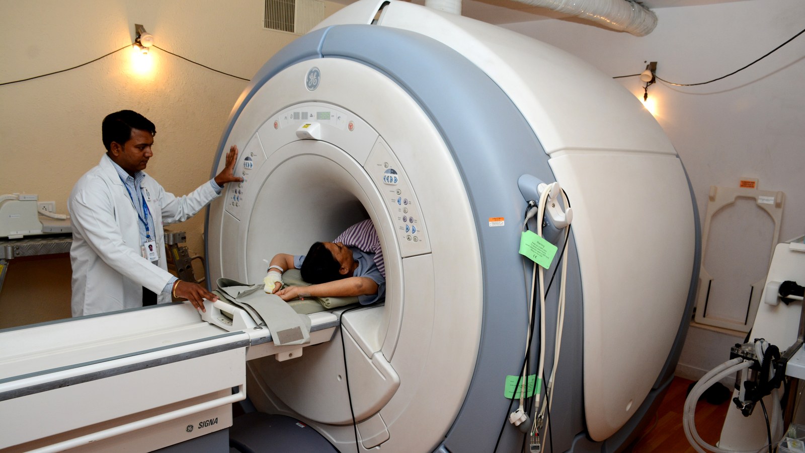
Image: clinical GE Signa MRI scanner / Wikimedia Commons. Does not show hProCA32.Collagen. Cropped to 16:9.
Source consulted: New MRI agent detects lung cancer | EurekAlert; Protein-based MRI agent | MedicalXpress.








0 Comments
No comments on this article yet. Be the first!